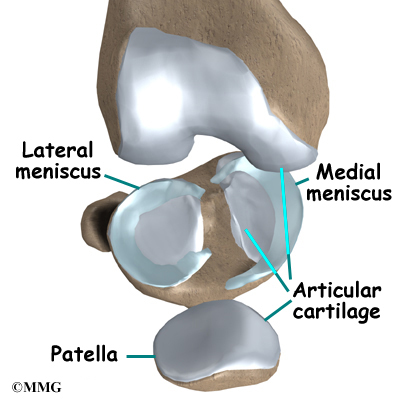
Knee Meniscus and ACL Surgery What You Need to Know Biology Diagrams The knee meniscus is a special layer of cartilage that lines the knee joint. The job of the meniscus is to cushion the knee joint and transfer forces between the tibia and femur, the thigh and shin bones. Most of the joints in our body are lined with a thin layer of articular cartilage, made of collagen and chondroitin. This provides a smooth Structure and Function of the Knee Meniscus 2.1 Meniscus Anatomy. The knee joint contains the meniscus structure, Transepiphyseal replacement of the anterior cruciate ligament in skeletally immature patients. A preliminary report. J Bone Jt Surg Am. 2003;85-A:1255-63. doi: 10.2106/00004623-200307000-00011.

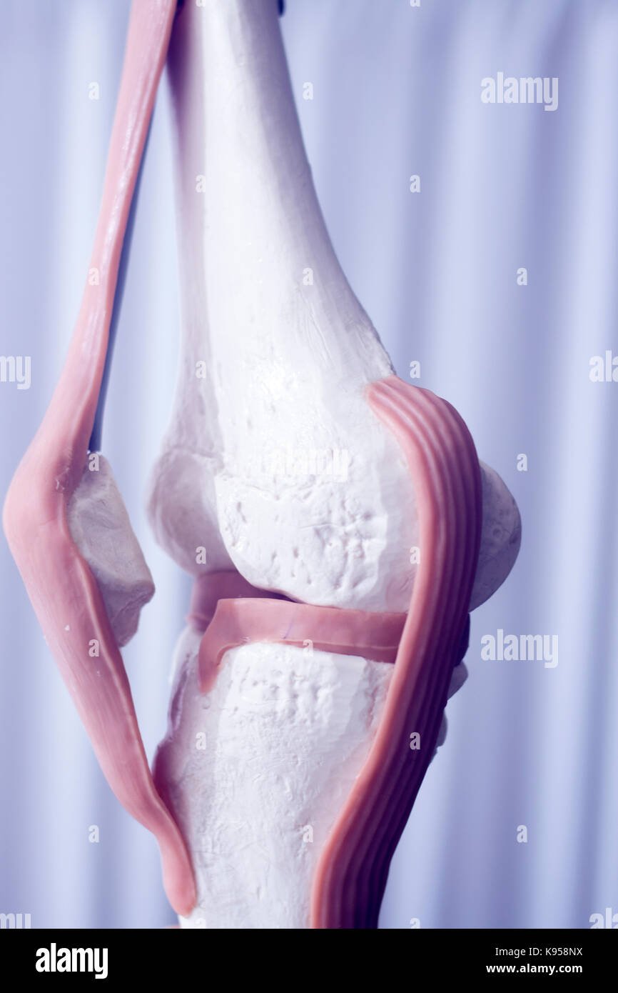
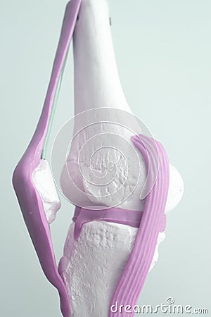
Information about the anatomy of the knee which connects the shinbones to the femur with the knee cap (patella), quadricpes muscles, hamstring muscles, anterior cruciate ligament (ACL), posterior cruciate ligament (PCL), medial collateral ligament(MCL) and lateral collateral ligament (LCL), lateral meniscus, medial meniscus
SHELBOURNE KNEE CENTER Biology Diagrams
The anterior cruciate ligament (ACL), is the weaker of two cruciate ligaments of the knee, the other being the posterior cruciate ligament.These intracapsular ligaments are so named due to the fact that they cross each other, creating an imaginary cross (the word cruciate comes from the latin word crux that means cross).
Anatomy. Gross Shape. medial meniscus. C-shaped with triangular cross section. to 10mm. average thickness of 3 to 5mm. lateral meniscus. is more circular (the horns are closer together and approximate the ACL) covers a larger portion of the articular surface. average width is 10 to 12mm. average thickness is 4 to 5mm. Knee & Sports
Knee Meniscus: Function & Injuries Biology Diagrams
Information about the anatomy of the knee which connects the shinbones to the femur with the knee cap (patella), quadricpes muscles, hamstring muscles, anterior cruciate ligament (ACL), posterior cruciate ligament (PCL), medial collateral ligament(MCL) and lateral collateral ligament (LCL), lateral meniscus, medial meniscus
