Intervertebral Disc Herniation Biology Diagrams Intervertebral Discs in Spine Anatomy. Intervertebral discs are cushion-like pads located between the bones of the spine (vertebrae). They are flat, round, and about half an inch thick. These discs have two main parts: Nucleus Pulposus: This is the jelly-like center of the disc. It contains much water, which gives the disc flexibility and helps Over time, spinal discs dehydrate and become stiffer, causing the disc to be less able to adjust to compression. While this is a natural aging process, as the disc degenerates in some individuals, it can become painful.. The most likely reason for this pain is that the degeneration can produce micromotion instability and the inflammatory proteins (the soft inner core of the disc) probably leak
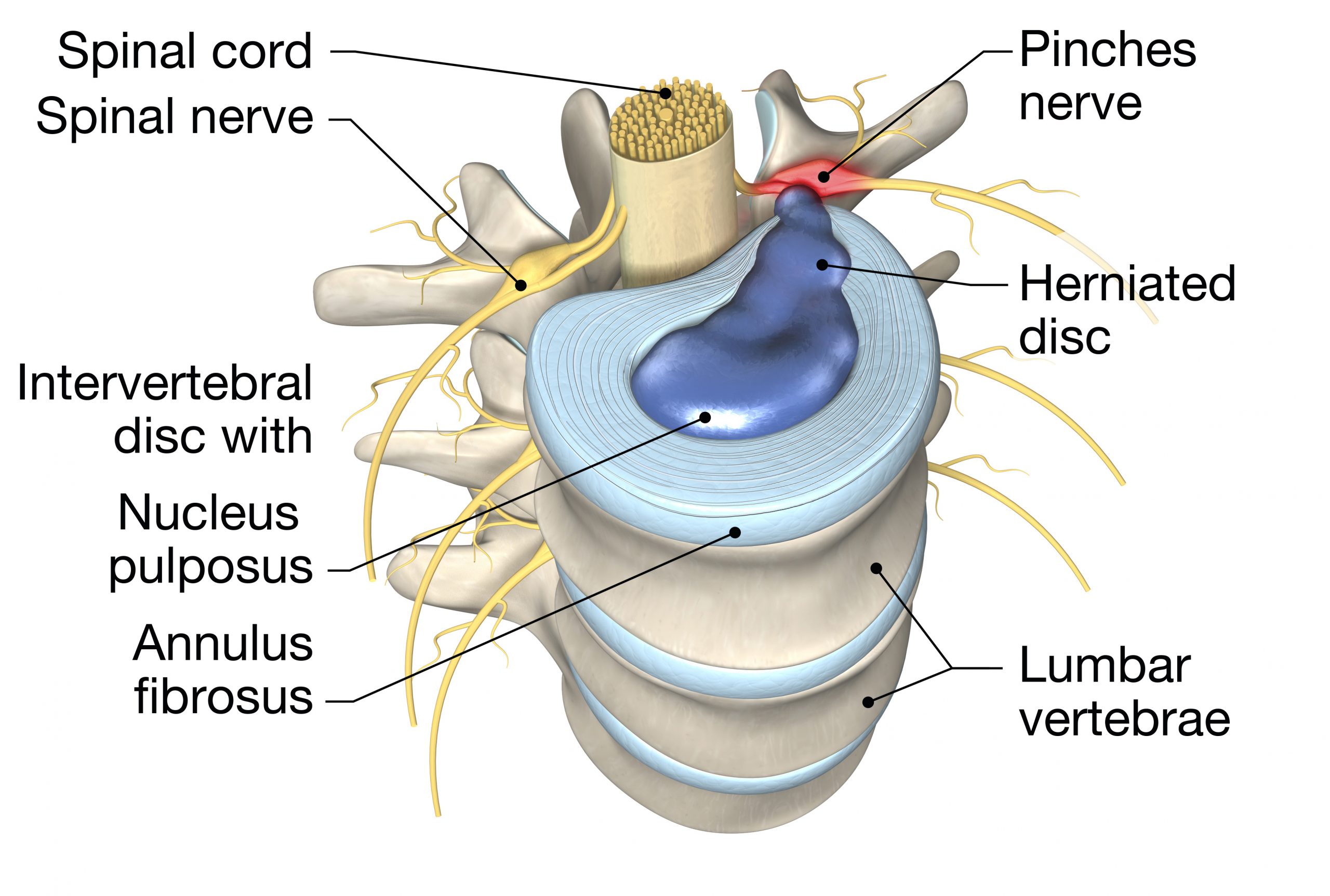
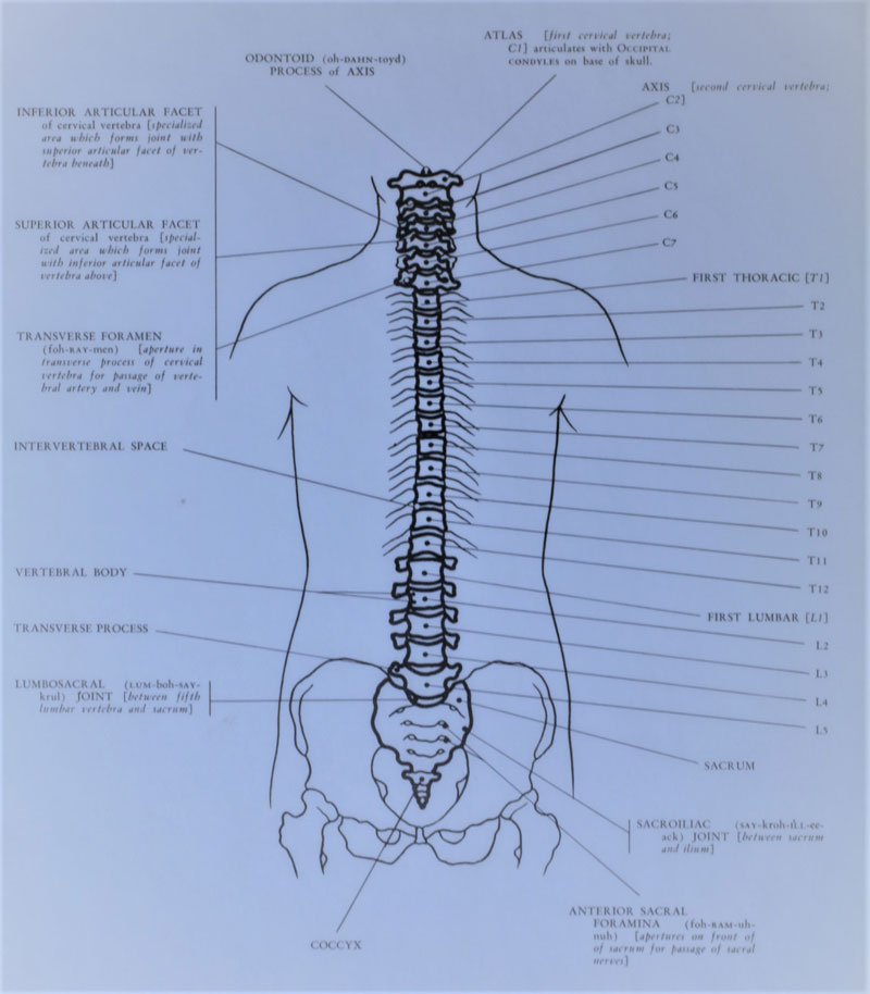
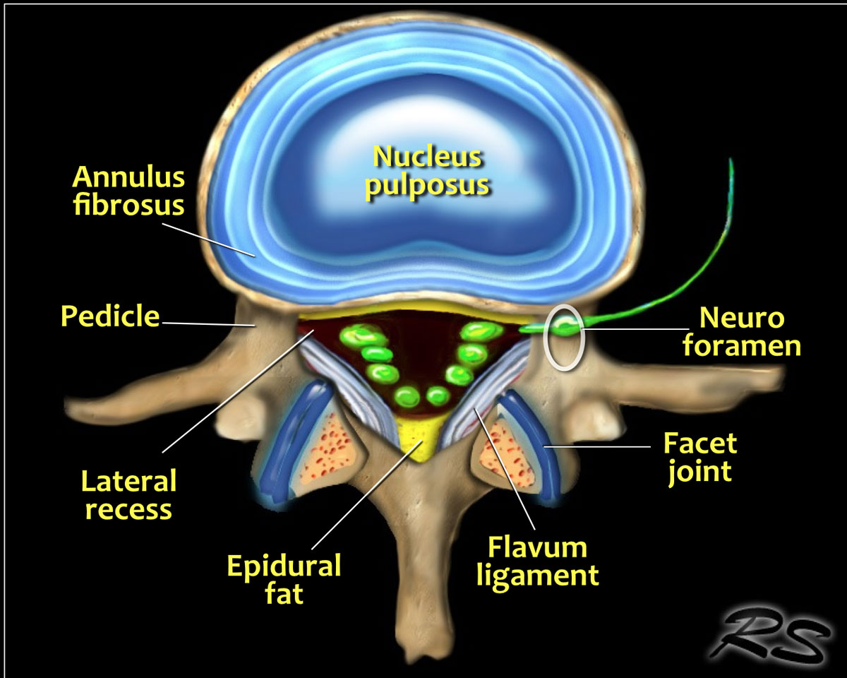
The intervertebral disc (IVD) is important in the normal functioning of the spine. It is a cushion of fibrocartilage and the principal joint between two vertebrae in the spinal column. There are 23 discs in the human spine: 6 in the cervical region (neck), 12 in the thoracic region (middle back), and 5 in the lumbar region (lower back). Anatomy of a Spinal Disc. Delving into the structure of a spinal disc, it becomes apparent that these complex components are comprised of two main parts: the outer annulus fibrosus and the inner nucleus pulposus. The annulus fibrosus is a tough, fibrous ring that provides strength and flexibility to the disc. It is primarily composed of type I

Intervertebral Discs: Structure, Function, and Disorders Biology Diagrams
These 25 discs (7 cervical, 12 thoracic, 5 lumbar, and 1 sacral) account for about 25% to 33% of the length of the spine. They allow the spine to be flexible without sacrificing a great deal of strength. They also provide a shock-absorbing effect within the spine and prevent the vertebrae from grinding together. This review article describes anatomy, physiology, pathophysiology and treatment of intervertebral disc. The intervertebral discs lie between the vertebral bodies, linking them together. The components of the disc are nucleus pulposus, annulus fibrosus and cartilagenous end-plates. The blood supply to the disc is only to the cartilagenous end
The inferior surface of the superior vertebral body articulates with the superior surface of the inferior vertebral body through intervertebral (IV) discs. These 25 discs (7 cervical, 12 thoracic, 5 lumbar, and 1 sacral) account for about 25% to 33% of the length of the spine. Intervertebral discs are fibrocartilaginous structures located between the bodies of adjacent vertebrae.They form a fibrocartilaginous joint between the vertebral bodies, linking them together. Collectively, the discs contribute up to one-third of the length of the vertebral column, forming an interpose between adjacent vertebrae from the axis (C2) to the sacrum.