Human Diaphragm Anatomy stock illustration Illustration of biology Biology Diagrams Learn more about the diaphragm anatomy with our custom quiz which covers the anatomy, blood supply, innervation and function of the diaphragm! J.R. Humpherson et al.: Human Anatomy, Colour Atlas and Textbook, 5th Edition, Mosby Elsevier (2008). R. Drake, A.W. Vogl, A.W.M. Mitchell: Gray's Anatomy for Students, 3rd Edition, Churchil
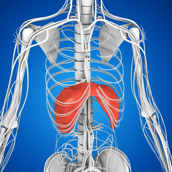
Structure of diaphragm shown using a 3D medical animation still shot. The thoracic diaphragm, or simply the diaphragm (/ ˈ d aɪ ə f r æ m /; [1] Ancient Greek: διάφραγμα, romanized: diáphragma, lit. 'partition'), is a sheet of internal skeletal muscle [2] in humans and other mammals that extends across the bottom of the thoracic cavity.The diaphragm is the most important muscle Anatomy . The diaphragm is a fibrous muscle that runs between the chest and abdomen, separating these two large cavities. It is asymmetric, as its right dome is larger than the left dome. The diaphragm has openings that allow certain structures to span the chest and abdominal cavities.
Structure, Anatomy, Function, Diagram, Location Biology Diagrams
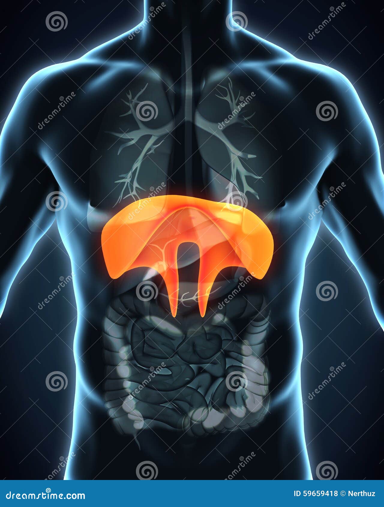
The muscles of the diaphragm arise from the lower part of the sternum (breastbone), the lower six ribs, and the lumbar (loin) vertebrae of the spine and are attached to a central membranous tendon. Contraction of the diaphragm increases the internal height of the thoracic cavity, thus lowering its internal pressure and causing inspiration of air. . Relaxation of the diaphragm and the natural The diaphragm in the thorax is called the thoracic diaphragm and serves as an important anatomical landmark that separates the thorax, or chest, from the abdomen. It functions during breathing when it contracts to enlarge the thoracic cavity and reduce the intrathoracic pressure so that lungs may expand and fill their alveoli with air. It is a dome-shaped muscle and tendon that functions as
The diaphragm separates the thoracic cavity from the abdominal cavity. It is a double-domed, musculotendinous partition that consists of a continuous sheet of muscle surrounding a central tendon. The peripheral muscle is named according to its peripheral points of attachment. The sternal part attaches to the posterior aspect of the xiphoid process. The coastal part attaches to the internal

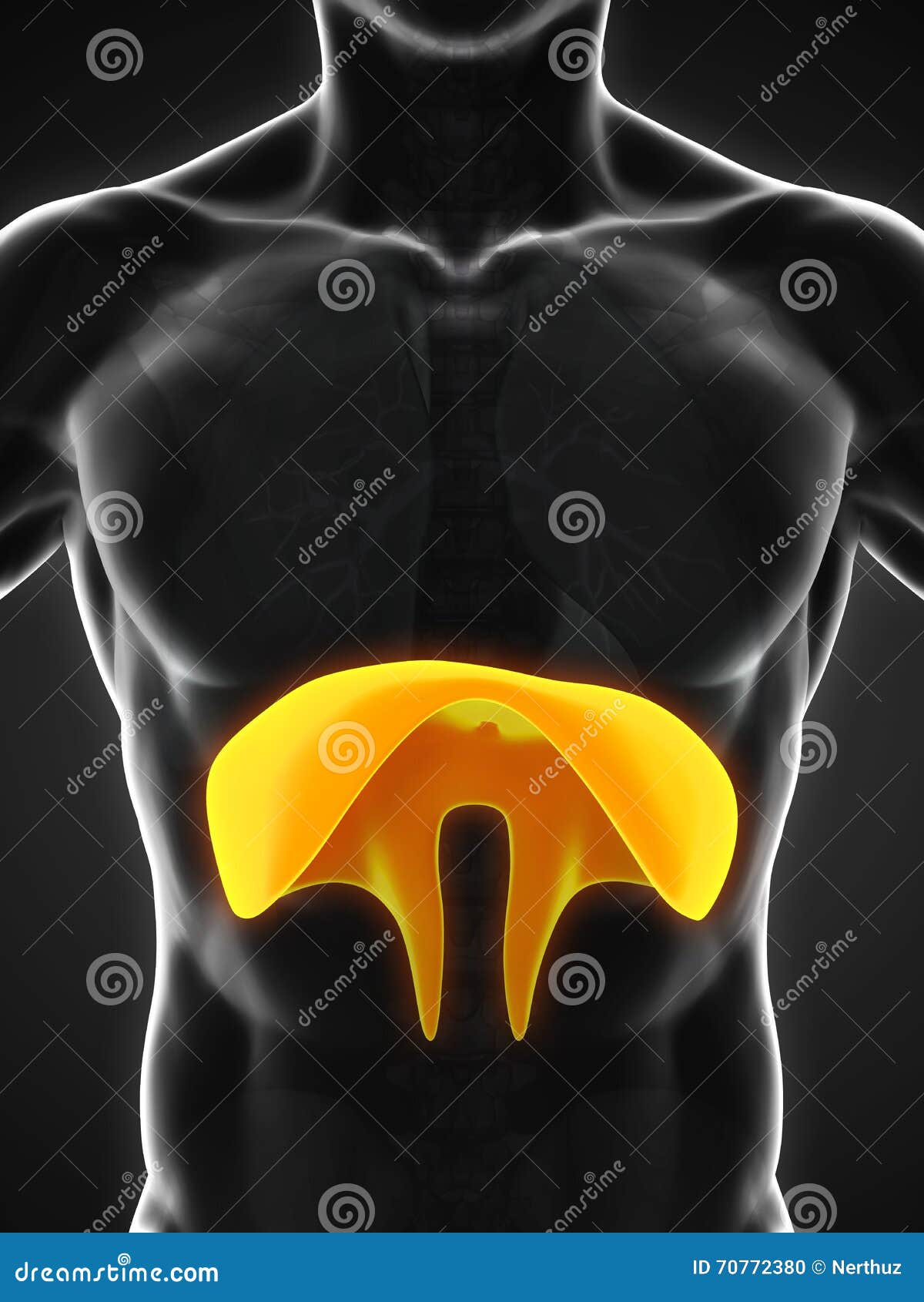
Diaphragm: Origin, Insertion, Openings, Function, Diagram Biology Diagrams
Della Barnes, an MS Anatomy graduate, blends medical research with accessible writing, simplifying complex anatomy for a better understanding and appreciation of human anatomy. What is the Diaphragm The diaphragm is a large, flat, double-domed sheet of muscle located in the thoracic region of the torso or body trunk.
