Anatomical Charts Posters Biology Diagrams A pleura is a serous membrane that folds back on itself to form a two-layered membranous pleural sac. The outer layer is called the parietal pleura and attaches to the chest wall. The inner layer is called the visceral pleura and covers the lungs, blood vessels, nerves, and bronchi. There is no anatomical connection between the right and left pleural cavities.[1] See Image. Lung Anatomy. With
A hollow area (pleural space) lies between the layers of your pleura. Your pleural space contains a thin layer of pleural fluid. Pleural fluid helps your pleura move as you breathe. The pleura has two layers: Visceral pleura. This is the inner layer of your pleura. It covers your lungs, blood vessels, bronchi and nerves. The visceral pleura
Anatomy, Thorax, Pleurae Biology Diagrams
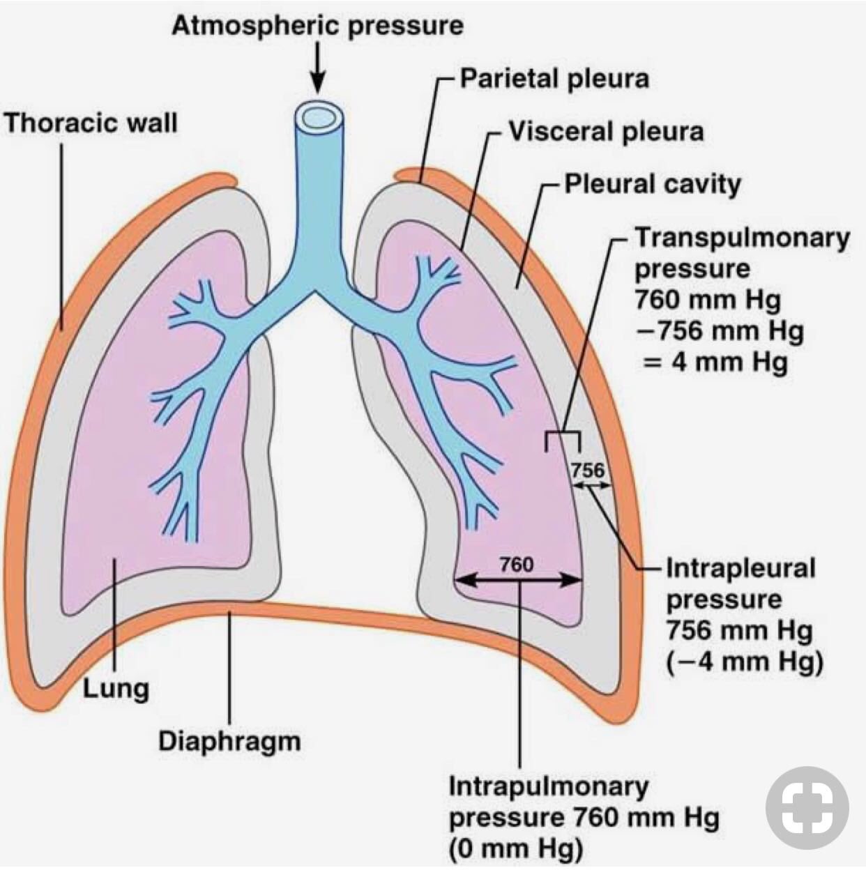
The pleura is a double-layered serous membrane that surrounds the lungs and lines the thoracic cavity. It provides a protective covering for the lungs and facilitates smooth respiratory movements. The pleura is divided into two layers—parietal pleura and visceral pleura—which are separated by the pleural cavity filled with a thin layer of pleural fluid. Pleural effusions. The excessive accumulation of fluid in the pleural cavity is called pleural effusion. It is usually due to inflammation of the pleura. The progressive accumulation of fluid may cause retraction of lung towards the hilum and may displace the mediastinum to the opposite side. The fluid may be in the form of: The pleural cavity is a fluid filled space that surrounds the lungs.It is found in the thorax, separating the lungs from its surrounding structures such as the thoracic cage and intercostal spaces, the mediastinum and the diaphragm.The pleural cavity is bounded by a double layered serous membrane called pleura.. Pleura is formed by an inner visceral pleura and an outer parietal layer.
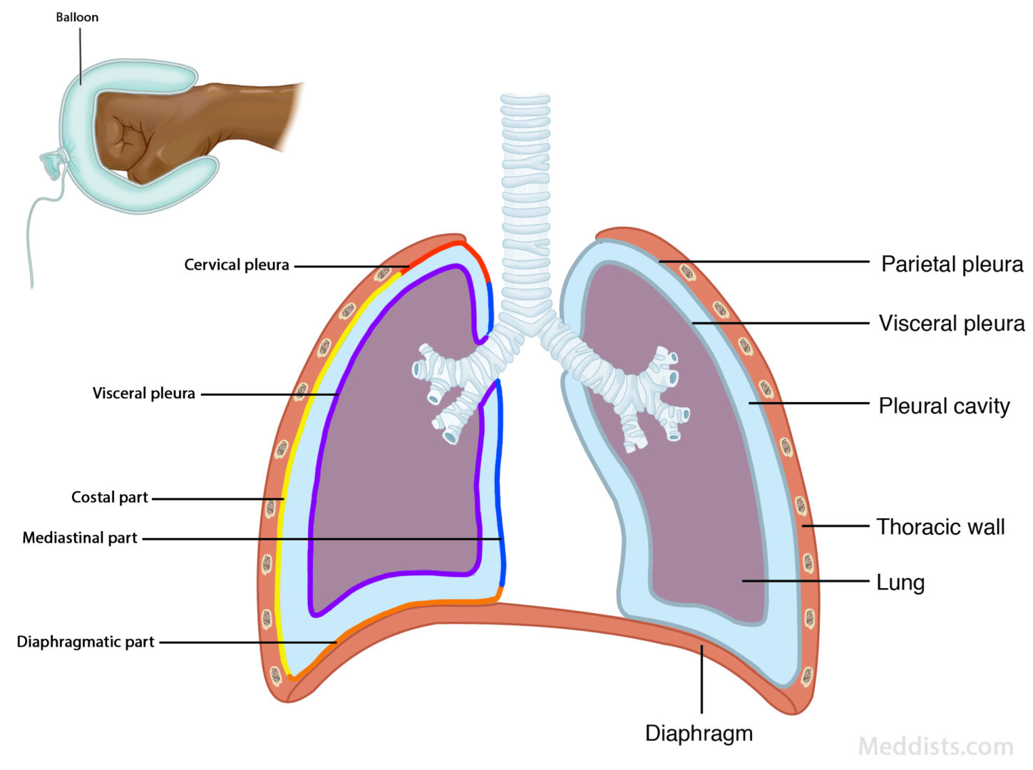
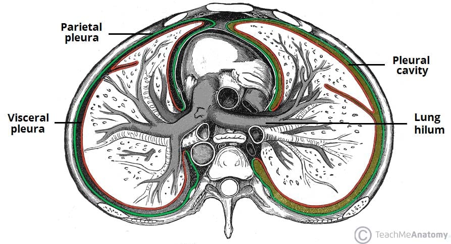
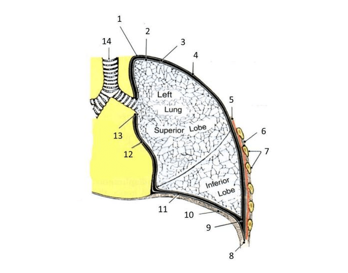
Each pleura can be divided into two parts: Visceral pleura - covers the lungs. Parietal pleura - covers the internal surface of the thoracic cavity. These two parts are continuous with each other at the hilum of each lung. There is a potential space between the viscera and parietal pleura, known as the pleural cavity. The pleura is a double-layered serous membrane that covers each lung and lines the thoracic cage.The outer layer (parietal pleura) attaches to the chest wall.The inner layer (visceral pleura) covers the lungs, neurovascular structures of the mediastinum and the bronchi.The space between the parietal and visceral pleurae is called the pleural cavity which contains a small amount of serous fluid
TeachMeAnatomy Biology Diagrams
Gross anatomy. The pleura divides into: visceral pleura: covers the surface of the lung and dips into the fissures between its lobes. lines of pleural reflection outline where parietal pleura abruptly changes direction as it passes from one wall of the pleural cavity to another. Right and left parietal pleura reflect in an asymmetric manner
