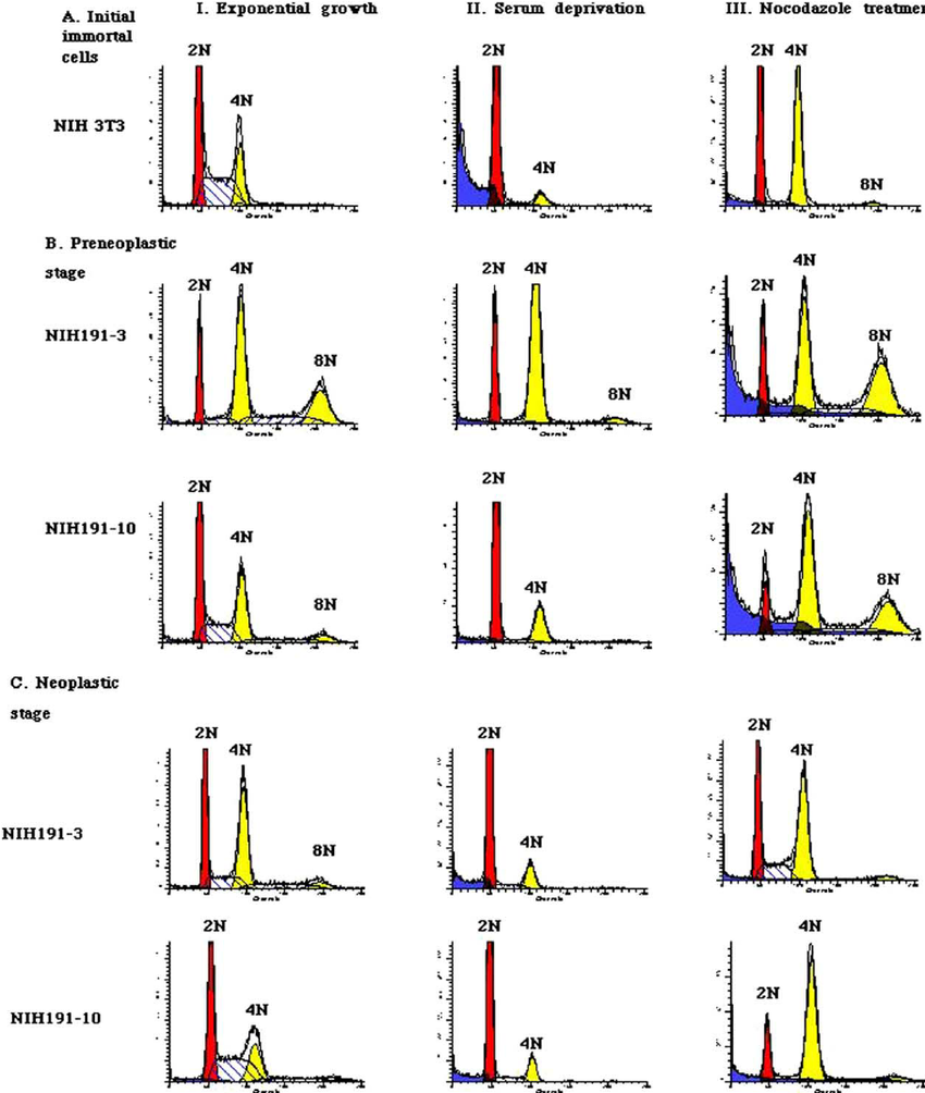
A Flow Cytometry of the cell cycle when the concentrations were 10 Biology Diagrams Figure 2. Cell cycle analysis with FxCycle Violet Ready Flow Reagent and Invitrogen Click-iT EdU Alexa Fluor 647 Flow Cytometry Assay Kit. Jurkat cells, a human T cell leukemia cell line, were pulsed with 10 µM EdU for 2 hours prior to detection with Alexa Fluor 647 azide.

Cell cycle analysis by quantitation of DNA content was one of the earliest applications of flow cytometry. The DNA of mammalian, yeast, plant or bacterial cells can be stained by a variety of DNA binding dyes. The premise of these dyes is that they are stoichiometric, i.e. they bind in proportion to the amount of DNA present in the cell. Abstract. In this unit, we describe two protocols for analyzing cell cycle status using flow cytometry. The first is based on the simultaneous analysis of proliferation specific marker (Ki-67) and cellular DNA content, which discriminates resting/quiescent cell populations (G0 cell) and quantifies cell cycle distribution (G1, S or G2/M, respectively).

Assaying cell cycle status using flow cytometry Biology Diagrams
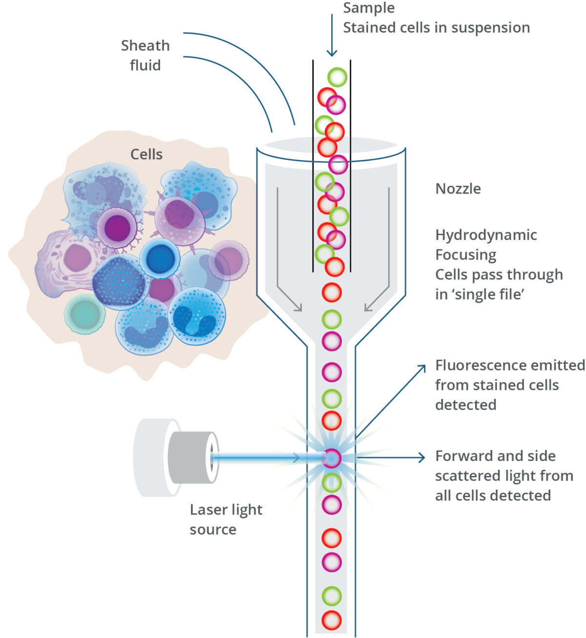
The ability to analyze thousands of cells per second makes flow cytometry invaluable for research and clinical applications. Phases Of The Cell Cycle. The cell cycle stages—G0/G1, S, and G2/M—are crucial for cell growth and division. Flow cytometry is particularly useful in analyzing these phases, offering insights into cellular dynamics. G0/G1 can be counterstained. Flow cytometry then allows the simultaneous measurement of incorporated BrdUrd as well as the DNA content on a single cell level. In this way the cohort of labeled cells can be followed through the cell cycle. Figure 2. DNA and cell cycle analysis. An example of DNA and cell cycle analysis.

Cell cycle analysis is one of the earliest applications in flow cytometry and continues to be highly used to this day. Since the first reported method of Feulgen-DNA staining, cell cycle analysis has continued to grow and mature. With the recent advances in DNA dyes, understanding of additional cell … Novel Fluorescence Probes for Sensitive Detection of Exogenous and Endogenous Nitric Oxide in Live Cells NAD+ Metabolism " A Link to Age-Associated Pathologies Practical Guide for Live Cell Cycle Analysis in Flow Cytometry Intracellular pH Measurement with Dual Excitation Fluorescence Sensor BCFL Novel Esterase Substrate for the Quantification
Analysis of Cell Cycle by Flow Cytometry Biology Diagrams
Cell suspension: Cells must be in a single cell suspension prior to any fixation and staining for cell cycle analysis as aggregates will impact your data interpretation. While both the cytometric and data analysis procedures to evaluate results have mechanisms to exclude aggregates, no method for eliminating them is perfect.